
Imaging/Radiology


Medical Radiology
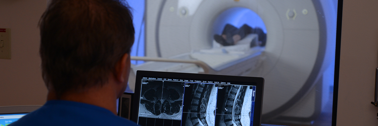
Medical Radiology is a term for X-rays, CT, MR and Nuclear Medicine exams as well as mammograms, ultrasound, and other tests and procedures that doctors request to help make a more accurate diagnosis.
Imaging and Radiology services at Hannibal Regional Hospital bring this advanced technology conveniently to the residents of northeast Missouri and west central Illinois. A variety of services and technologies are available to help doctors make the best, most informed diagnoses.
Our well trained staff is here to take care of your healthcare needs. To schedule an appointment in the Medical Radiology department, please call 573-248-5688.
Hannibal Regional Radiology/Medical Imaging Services
Hannibal Mobile Diagnostics
HMD Mobile Diagnostics provides mobile X-Ray and EKG services to NE Missouri and West Central Illinois nursing homes, retirement communities and correctional facilities. We come to you! This allows your residents to stay in the comfort of their own surroundings.
Hours of Operation:
- Monday-Friday 7am-10pm
- Saturday & Sunday 8am- 5pm
To Order an Exam:
- Fax HMD’s mobile order form (provided to the facility) to 888-851-5923 or log onto the physician portal (once you have registered and been given access) and place the order.
- Call 573-248-5123 to ensure the fax/order was received.
- Our Radiology Technologist will come to your facility to perform the services.
- Once the radiologist has read the exam, a report will be faxed to your facility and to the ordering physician.
Physician Portal Mobile: https://rad.hannibalregional.org/NewPhysicianPortalMobile/default.aspx
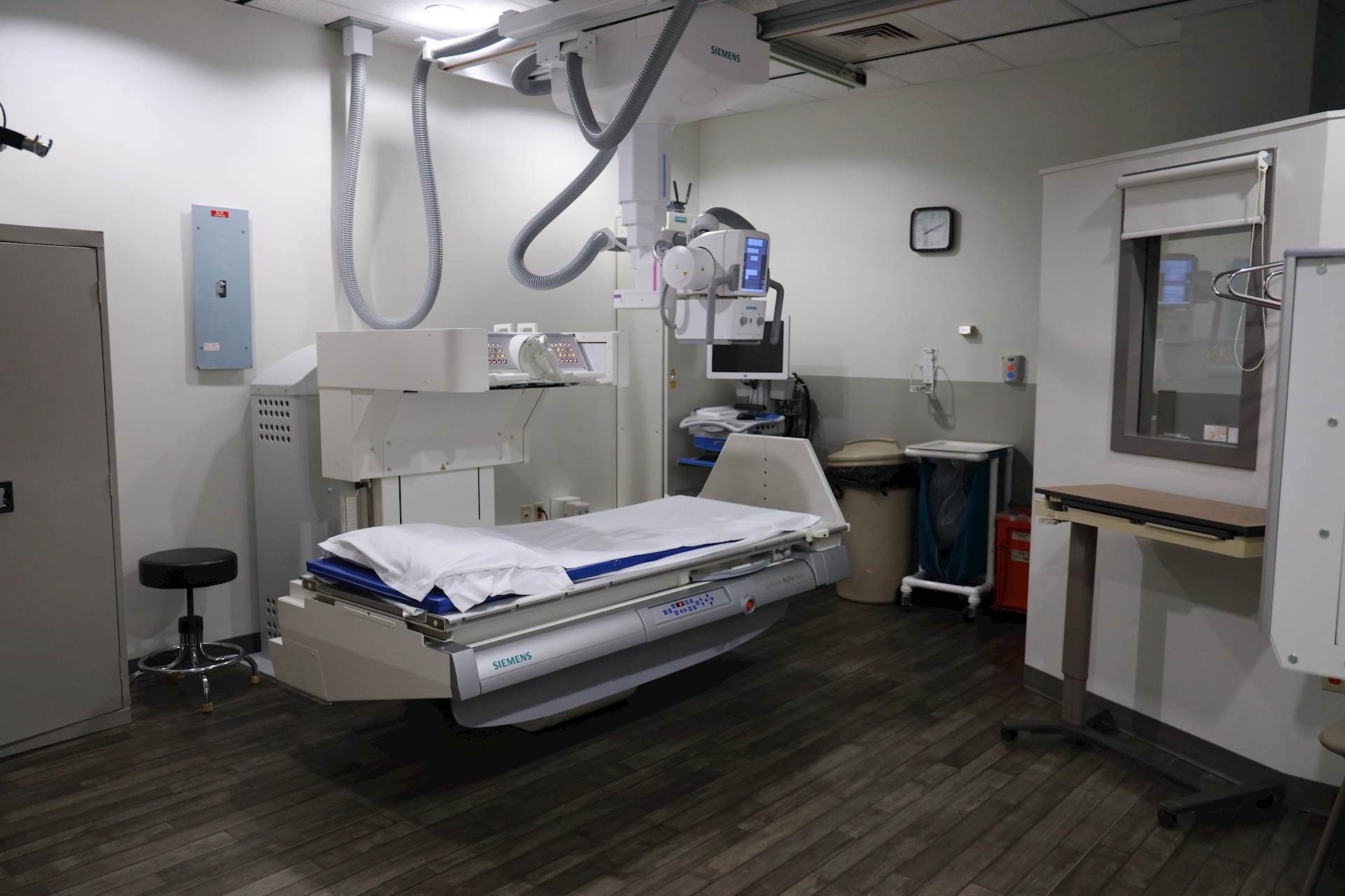
General Radiography/Fluoroscopy
We understand that our NE Missouri and West Central Illinois patients seek a thorough and caring approach and getting you in and out as quickly as possible is our priority.
Type of X-Ray Imaging scans we provide:
- General Radiographic (X-Ray)
- Fluoroscopic:
- Modified Barium Swallows
- Esophogram
- Upper GI
- Small Bowel Series
- Barium Enemas
- Hysterosalpingogram (HSG)
- Voiding Cystourethrogram VCUG
- Lumbar Punctures
- Myelograms
- Arthrograms
- Joint Aspiration/Injection
- Dobhoff Placement
Outpatient Hours of Operation:
- Hannibal Regional Hospital
- Monday- Friday 07:00am -5:00pm
- Hannibal Regional Medical Group- Bowling Green (General Radiographic/X-Ray Services Only)
- Monday- Friday: 08:00am -5:30pm
- Saturday and Sunday: 08:00am -4:00pm
- Hannibal Regional Medical Group- Canton (General Radiographic/X-Ray Services Only)
- Monday- Friday: 08:00am -5:00pm
- Hannibal Regional Medical Group- Hannibal (General Radiographic/X-Ray Services Only)
- Monday- Friday: 08:30am -5:00pm
- Hannibal Regional Medical Group- Monroe City (General Radiographic/X-Ray Services Only)
- Monday, Tuesday, Thursday and Friday: 08:00am- 12:00pm
- Wednesday: 01:00pm to 4:30pm
- Hannibal Regional Medical Group- Shelbina (General Radiographic/X-Ray Services Only)
- Monday, Tuesday, Thursday and Friday: 01:00pm to 4:30pm
- Wednesday: 08:00am- 12:00pm
- Complete Family Medicine- Kirksville- (General Radiographic/X-Ray Services Only)
- Sunday-Saturday: 08:00am-8:00pm
Preparation instructions for procedures:
- Appointments are not required for General Radiographic (X-Ray).
- Tell us if you are, or suspect you might be pregnant.
- Upper GI and small bowel series: nothing to eat or drink 8 hours before the exam.
- Barium Enemas: Will need to pick up colon prep from Hospital Team Member Pharmacy (Hours: Monday-Friday 0730- 4:30pm) At least 3 days prior to exam. Detailed instructions will be given at time of prep pick up.
- Hysterosalpingogram: requires a negative pregnancy test before starting the exam.
- Lumbar punctures and Myelograms: A Radiology Nurse will reach out to the patient one business day before procedure with specific instructions.
Women's Imaging
Hannibal Regional’s Women's Imaging has a state-of-the-art, 3D-Tomosynthesis Digital Mammography unit, in addition to Digital Computer Assisted Technology (CAD). 3D mammography or tomosynthesis has the ability to detect subtle findings while minimizing callbacks for additional testing. This unit is available in both screening and diagnostic mammography studies.
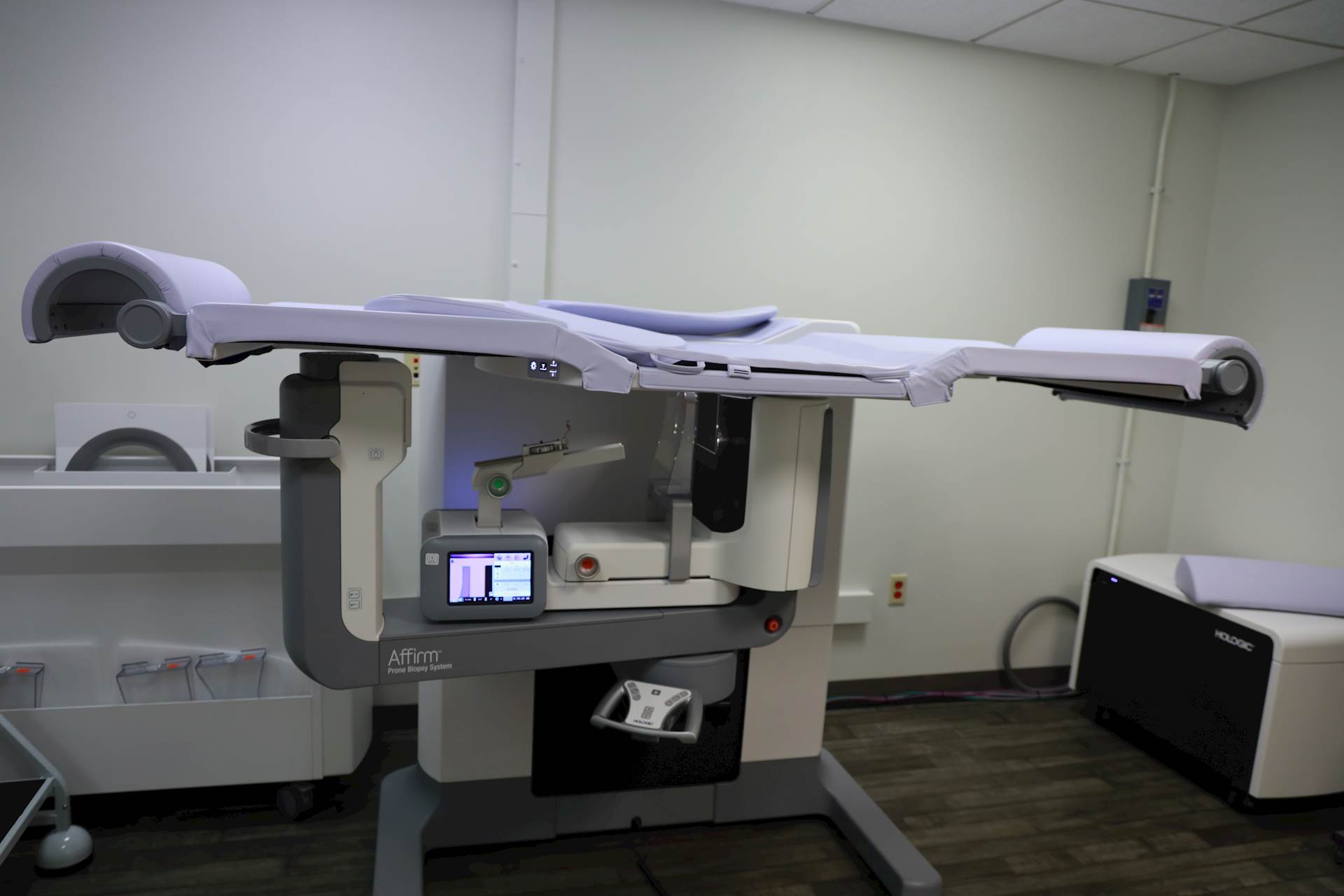
A state of the art stereotactic procedure room with capabilities for 3D localization allows for additional diagnostic technology and minimally invasive procedures to diagnose breast abnormalities.
Hannibal Regional’s Women's Imaging is a comprehensive diagnostic department that is designed to offer rapid response to the needs of the patients. Arrangements are made to meet each patient's medical needs in a timely and effective manner. Under the care of a breast health navigator, this dedicated, individualized service makes it possible for the patient's diagnostic workup to begin as soon as possible.
Screening exams available:
- Mammogram (patients do not need a doctor's order) 2D or 3D-Tomosynthesis.
- Ultrasound and breast MRI (at the discretion of your doctor).
- Bone density scans.
If further testing is needed, we also can perform the following tests:
- Diagnostic mammogram (requires a doctor's order) 2D or 3D-Tomosynthesis.
- Diagnostic ultrasound.
- Breast MRI
- MRI-guided breast biopsy
- Stereotactic and 3D guided breast biopsy
- Ultrasound-guided biopsy
- Needle Localization
Outpatient Hours of Operation:
- Hannibal Regional Hospital
- Monday- Friday 07:30am -4:45pm
Preparation instructions for procedure:
Mammography:
Avoid using deodorants, antiperspirants, powders, lotion and creams under the arms or breasts to avoid any metallic particles from showing up on the mammogram from these products
If you have not entered menopause, schedule your mammogram for a week following your menstrual period to have less tender breasts and consider using an over the counter pain reliever one hour prior to the mammogram to reduce any discomfort of the test.
DEXA (bone density):
Do not take calcium supplements or anything containing a large dose of calcium such as antacids 24 hours prior to the test. These might show up on the test giving a false result if they are not entirely dissolved. wear loose fitting clothing that is free of metal zippers and buttons.
Stereotactic:
If you are prescribed Warfarin (Coumadin), you will need to have a blood test 3 days prior to the biopsy to check you blood clotting factors. Bring a list of your allergies to the test. Wear a tight bra such as a sports bra to the exam as this will help hold compression and the small ice pack that is given after the biopsy.

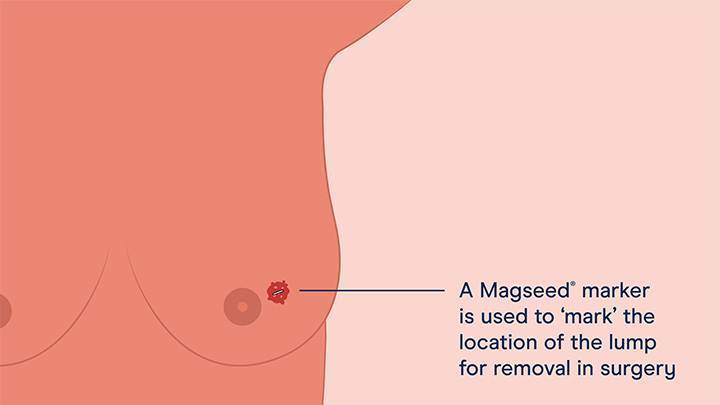
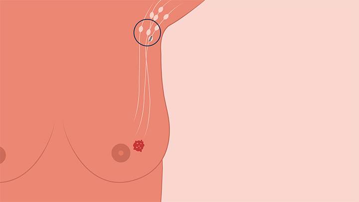
Magseed® Marker
In order for the surgeon to know the exact location of the tumor, there are a number of aids that can be used to ‘mark’ it. One of these is the Magseed marker. The Magseed marker will be placed in the center of your tumor in advance of your surgery. Once you are in the operating room, your surgeon will first use your mammogram images to give them a rough idea of where the tumor is located in your breast. A probe will then be used to detect the signal from the Magseed marker, which will enable them to accurately locate the seed. This enables the tumor to be removed accurately and completely.
The Procedure - What to expect:
- Your appointment in radiology
With a ‘Magseed localization’ procedure, you will attend a placement appointment prior to your surgery.
Your Magseed marker could be placed many weeks before your planned surgery date, at a time that suits both you and your surgeon.
When it’s time to place the marker, a local anesthetic will be injected where the marker will be placed. Your radiologist will then use an ultrasound machine (or another imaging approach) to identify the area of suspicion found on your mammogram.
They will then insert the Magseed into the area, ensuring the seed is placed accurately and securely. This is a quick and efficient procedure.
After your Magseed has been inserted, you’ll be able to carry on life as normal up to your date of surgery, without fear of discomfort or displacement. - Removing the seed during surgery
The surgery to remove a tumor containing a Magseed marker is a routine procedure for a breast surgeon.
With your Magseed already placed, you will be ready to go straight through to the operating room at the agreed appointment time.
During the surgery, your surgeon will use a probe to detect and locate the previously placed Magseed marker in you breast - with the marker's magnetic signal accurately guiding them to the site of the cancer to be removed. - What happens after the surgery?
Once the tumor has been removed, it will be sent to pathology for closer examination, to determine whether the cancer has been completely removed.
The likelihood of leaving any of the tumor behind with the Magseed marker has been shown to be very low.
Magtrace® Lymphatic Tracer
To help identify the sentinel nodes, the Magtrace lymphatic tracer will be injected into your breast. This magnetically detectable ‘tracer’ liquid will follow the route that a migrating cancer cell would take, before collecting in the sentinel nodes in your underarm.
Once the tracer has migrated to your sentinel nodes, your surgeon will then be able to identify the location of these nodes by using a magnetic probe which can detect even minute quantities of the tracer liquid.
The unique properties of Magtrace identifies the nodes most likely to contain cancer on the basis of their position in the lymphatic chain thereby minimizing the number of nodes needed to assess the local extent (spread) of the cancer.
The Magtrace Procedure
- The injection before surgery
The Magtrace® lymphatic tracer will be injected at a time that suits you and your surgeon.
It can be administered at the start of your surgery when you’re under anesthesia, or many days or weeks beforehand, timed to coincide with your pre-surgical visit to the hospital or at a time that is convenient to you.
If administered beforehand, local anesthetic or a numbing gel will be applied. Your surgeon will then use a small needle to inject the liquid into your breast. - After the injection
Shortly after the injection, the Magtrace lymphatic tracer liquid will flow through your lymphatic system, taking the route a migrating cancer cell would take, before collecting in the sentinel nodes in your underarm.
Only the sentinel lymph nodes will be marked by Magtrace.
In a small number of patients injected with Magtrace, discoloration of the skin at the injection site can occur.
This is a tiny amount of the tracer which has remained under the skin. Any discoloration will fade over time. - The surgery
During the procedure, your surgeon will use a Sentimag probe to locate the Magtrace lymphatic tracer in your sentinel lymph nodes. Once found, your surgeon will usually remove two or three nodes for analysis.
A pathologist will examine the specimens to confirm if any cancer cells are present in the lymph nodes and help determine if the cancer has begun to spread.
This process can take some time, so if you do not hear back straight after surgery, do not worry.
What will the results from the analysis tell me?
The analysis will determine if cancer cells are present in your lymph nodes and how much is in each one.
This will help to accurately stage your cancer and help determine your best course of treatment, which may include additional therapy, such as chemotherapy and radiation.
Ultrasound (US)
Ultrasound is a safe non-invasive, no radiation exam performed on most parts of the body for various purposes. Advanced Imaging at Hannibal Regional utilizes a fleet of Philips Epiq 7 machines, capable of producing high quality diagnostic images.
Exams:
- Echocardiogram
- Abdominal
- Pelvic
- Carotid
- Thyroid
- Vascular
- Breast
- Gynecological
- Obstetric
- Infant echo/spine/heads
- Pediatric
- Testicular
- Ultrasound guided biopsies
- Paracentesis/Thoracentesis
Outpatient Hours of Operation:
- Hannibal Regional Hospital
- Monday- Friday 7:00am to 3:30pm
- HRMG OB/GYN-Hannibal (OB/GYN Services)
- Monday- Friday 7:00am to 4:30pm
Preparation instructions for procedure:
- On the day of your ultrasound, it is recommended that you wear a lose-fitting, two-piece outfit.
- Female patients may be asked to drink water to fill their bladders prior to certain exams.
- A Sonographer or technologist will reach out to patients who will need to be NPO (nothing to eat or drink) for their procedure.
- If you are having an ultrasound interventional procedure, a radiology nurse will call you prior to your appointment to provide you with instructions.
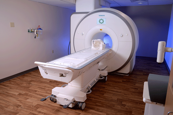
Magnetic Resonance Imaging (MRI)
Hannibal Regional’s MRI technology provides our patients with a much more comfortable imaging experience and our physicians with more detailed scans and enhanced diagnostic confidence. The Siemens Magnetom Aera 1.5 Tesla MRI creates ultra-high-field, next generation imaging while the more spacious design reduces anxiety and its speed means faster exam times.
Speaking of design, the Siemens Aera 1.5 Tesla offers a bore design which is more spacious. This design helps reduce the closed-in feeling of traditional MRIs. This helps reduce anxiety and its speed means faster exams, resulting in a more comfortable experience for you.
Type of MRI scans we provide:
- General magnetic resonance imaging (MRI) Scans
- MRCP
- MRI breast
- MRI guided breast biopsies
- MRI Enterography
- Magnetic resonance angiography (MRA)
Outpatient Hours of Operation:
- Hannibal Regional Hospital
- Monday- Friday 08:00am -4:00pm
Preparation instructions for procedures:
- Exams with contrast requires laboratory work to check for creatinine and BUN blood levels prior to an IV contrast exam.
- Patients with pacemakers can be scanned with prior approval from Pacemaker vendor and proper assessment of pacemaker before and after the study.
- Patient with pain stimulators can be scanned with prior approval allowing stimulator to be placed at a specific MRI setting prior to the test.
- You will be asked to complete a questionnaire before your MRI exam. Because of the potential harmful effects associated with some metallic objects in a magnetic field, you should tell the technologist performing your exam if you have had any surgeries, or if you have a pacemaker, aneurysm clips, metal in your eyes, metal implants in your ears, an implanted drug infusion device, shrapnel or bullet wounds, or permanent eyeliner.
- If you have ever been a metal worker, you may be required to have special x-rays before your exam to make sure there are no metal fragments in your eye(s).
- You will be asked to remove all metallic items from your person, such as watches, jewelry, hairpins, eyeglasses, and hearing aids. Also, do not take credit, bank, or parking cards with you into the scanner - the magnet will erase the information recorded on the metallic strip. An area for the safekeeping of your valuables is provided outside the scanner area.
- Plan for at least 1 hour to complete each MRI examination. The length of your scan will depend on the type of information needed and may require more or less time.
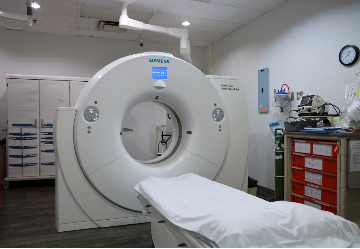
Computed Tomography (CT)
Hannibal Regional offers state-of-the-art Computed Tomography (CT) scanners at Hannibal Regional Hospital and Hannibal Regional Medical Group in Bowling Green. Hannibal Regional’s equipment, uses innovative technologies to provide better imaging using less radiation and contrast.
CT Scanners offers many new features that allow for the lowest radiation dosage, offering exams that result in 80% lower radiation dose than previously seen. The large opening of the CT, coupled with much faster scan speeds (and lower breath-hold times for our patients), will provide a better experience for every patient who visits our department for a CT scan.
Type of CT scans we provide:
- General CT Scans
- CT Angiogram
- Cardiac CT Angiogram
- CT Coronary Calcium Scoring
- CT Virtual Colonography
- CT Biopsy
Outpatient Hours of Operation:
- Hannibal Regional Hospital
- Monday- Friday 07:00am -5:00pm
- Hannibal Regional Medical Group- Bowling Green (limited services based on insurance authorization or CT study).
- Monday- Friday: 08:00am -5:00pm
- Saturday and Sunday: 08:00am -4:00pm
Preparation instructions for procedures:
- Plan for at least 30 minutes to complete your CT examination. The length of your scan will depend on the type of information needed and may require more or less time.
- Patients older than 50 years old will need laboratory work to check for creatinine and BUN blood levels prior to an IV contrast exam.
- Cardiac Angios, biopsies, drain placements: A Radiology Nurse will reach out to the patient one business day before procedure with specific instructions.
- CT Virtual Colonography: Will need to pick up colon prep from Hospital Team Member Pharmacy (Hours: Monday-Friday 0730- 4:30pm) At least 3 days prior to exam. Detailed instructions will be given at time of prep pick up.
Interventional Radiology
Hannibal Regional performs a wide variety of complex diagnostic and interventional procedures. The idea behind interventional radiology is to diagnose and treat patients using the least invasive approach using imaging guidance to decrease risk to the patient and improve health outcomes. Interventional Radiology procedures have less risk, less pain and less recovery time compared to open surgery.
Interventional Radiology Procedures:
- Image guided breast biopsy: Stereotactic, ultrasound and MRI guided
- Arthrograms
- Lumbar punctures, blood patches, and myelograms
- CT, Flouro and US guided joint injections (knees,hips, shoulders, AC joint, wrist, ankle, SI joints)
- CT guided biopsies, aspirations, and drain placements
- Flouro and CT guided bone biopsies
- Bone marrow biopsy
- CT guided LESI
- US guided biopsies (Soft tissue, lymph node, and solid organ)
- US guided aspirations and drain placement
- Paracentesis
- Thoracentesis
- Pleurex catheter placement (chest and abdomen)
- Neural foramen injections
- Central venous access:
- Port placements (all types) (port removal offered)
- Tunneled Hemodialysis catheters (removal also offered)
- Non-tunneled hemodialysis catheters
- Tunneled PICC lines (Patients requiring IV access greater than six weeks or GFR <60)(removal offered)
- Non-tunneled PICC lines
- Central lines for difficult venous access by special request
- IVC filter placement and retrieval (we use retrievable filters)
- Trans jugular liver biopsy
- Percutaneous Cholangiograms and biliary drain placement
- Cholecystostomy tube placements
- Percutaneous nephrostomy tube placements (can also leave wire for urology)
- Antegrade ureteral stent placement
- Suprapubic catheter placement
Preparation instructions for procedures:
- Preparation instructions vary per procedures, therefore, a Radiology Nurse or Technologist will reach out to the patient one business day before procedure.
Advanced Interventional Radiology
Hannibal Regional performs a wide variety of complex diagnostic and interventional procedures. The idea behind interventional radiology is to diagnose and treat patients using the least invasive approach using imaging guidance to decrease risk to the patient and improve health outcomes. Interventional Radiology procedures have less risk, less pain and less recovery time compared to open surgery.
Advanced Interventional Radiology Procedures:
- Mechanical DVT thrombectomy (acute and chronic DVT)
- Bone tumor ablations
- Kyphoplasty/vertebroplasty ( tumor and fracture treatment)
Preparation instructions for procedures:
- Preparation instructions vary per procedures, therefore, a Radiology Nurse or Technologist will reach out to the patient one business day before procedure.
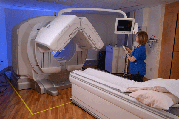
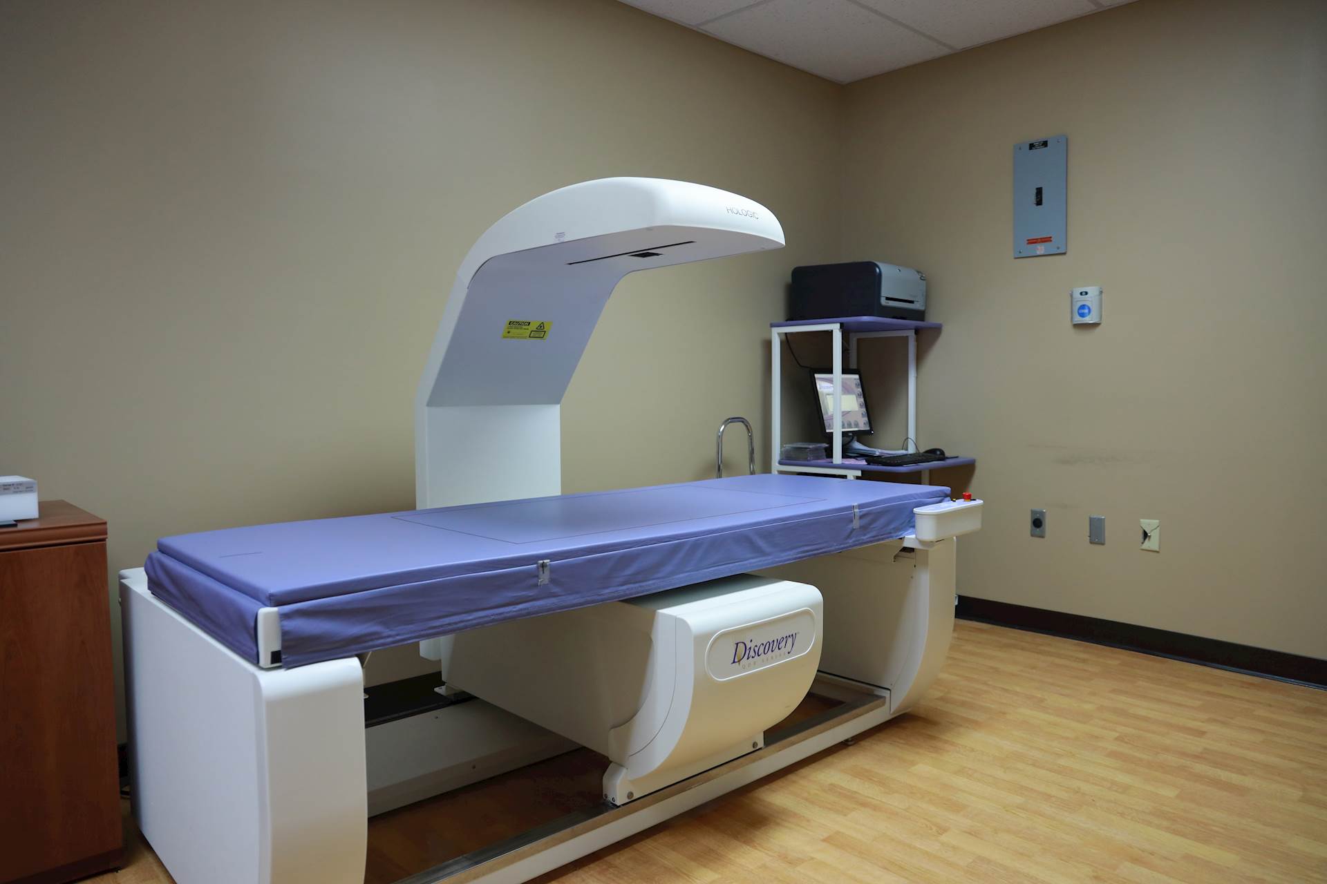
Nuclear Medicine
Hannibal Regional is excited to offer the latest in Nuclear Medicine imaging with the Discovery NM/CT 670. This unit is a hybrid Nuclear Medicine system which allows Nuclear Medicine images to be fused over 3D CT images to give our physicians precise diagnostic information and supports better patient treatment plans.
A nuclear medicine (NM) scan is a diagnostic exam that provides your doctor with important information about organ function. In nuclear medicine, imaging radiopharmaceuticals (radioactive medicine or tracers) are taken orally, by intravenous injection or inhalation. Special cameras then capture and form images from the radiation emitted by the radiopharmaceuticals in the body. This process is different from diagnostic X-rays which pass external radiation through the body to form the images.
Depending on the study, the procedure can range from 30 minutes to a few hours. In some cases, multiple day studies in which the patient leaves and returns at specific times are necessary. The radiation from the pharmaceutical is generally very minimal.
Types of Nuclear Medicine Scans we provide:
• Bone Scan
• Endocrine Scan
• Parathyroid Scan
• Lung Scan
• Cardiac Perfusion
• Gastrointestinal Scan
• Gall Bladder Scan
• Sentinel Lymph Node Identification
Outpatient Hours of Operation:
• Hannibal Regional Hospital
o Monday- Friday 07:00am -5:00pm
Preparation instructions for procedures:
• Preparation instructions vary per procedures, therefore, a Nuclear Medicine Technologist will reach out to the patient one business day before procedure.
Positron Emission Tomography (PET)
Our Imaging Providers
Click on the picture for more details.


